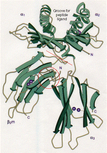
Schematic representation of the extracellular portion of HLA-A2, a human class I histocompatibility molecule. The stretches of beta conformation are represented by the broad green arrows (pointing N -> C terminal). Regions of alpha helix are shown as helical ribbons. The pairs of purple spheres represent the disulfide bridges.
The molecule of beta-2-microglobulin (β2m) is bound to the junction of the alpha1 (α1) and alpha2 (α2) domains (at the red line) and to the alpha3 domain (red circle) by noncovalent interactions only.
Not shown is the presence of a short peptide bound noncovalently in the groove between the alpha helices of the alpha1 and alpha2 domains. The combination of peptide and adjacent portions of the alpha helices makes up the epitope seen by CD8+ T cells.
| Link to a discussion of antigen presentation by class I histocompatibility molecules. |
The transmembrane and cytosolic portions of the molecule are also not shown.
(Courtesy of P. J. Bjorkman from Nature 329:506, 1987.)
| Welcome&Next Search |