| Link to discussion of the physiology of blood capillaries |
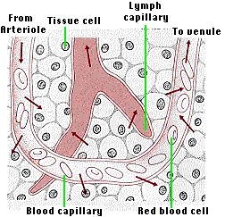
Most (~90%) of this interstitial fluid returns at the venule end of the capillary. The 10% that does not is picked up by tiny vessels called lymph capillaries. The cells forming the walls of the lymph capillaries are loosely fitted together, thus making the wall very porous. Any serum proteins that filtered through the blood capillary wall pass easily from the tissue space into the interior of the lymph capillary. (The lymph capillaries of the intestinal villi, called lacteals, also pick up fat droplets — link to discussion.) White blood cells (leukocytes) migrate from the tissue space into the lymph capillary squeezing between the cells that make up its wall.
The lymph capillaries drain into still larger collecting vessels.
The flow through the collecting vessels is quite slow. Like blood in the veins, contraction of skeletal muscles compresses the collecting vessels and squeezes the fluid — now called lymph — along. Again, like the return of blood in the veins, the lymph can flow only in one direction because of valves in the vessels.
All the lymph collected from| Link to drawing showing the anatomy of the lymphatic system (52K). |
The thoracic duct empties into the left subclavian vein. The lymph in the right side of the head, neck, and chest is collected by the right lymph duct and empties into the right subclavian vein. Together they empty 1–2 liters of lymph into the blood each day.
The collecting vessels of the lymphatic system are interrupted by lymph nodes.
These are especially abundant in theThese contain cavities — called sinuses — into which the lymph flows bringing various leukocytes (e.g., lymphocytes and dendritic cells) and out of which pass
which then enter the blood at the subclavian veins. [More]The walls of the sinuses are lined with phagocytic cells, which engulf any foreign particles, e.g., bacteria, that might be present in the lymph. Tests have demonstrated that over 99% of the bacteria carried into a node are screened out before the lymph leaves the node on its return to the blood.
This filtering mechanism is one of the most important body defenses against infectious disease. When combating a heavy infection, the lymph nodes enlarge producing "swollen glands".
| Welcome&Next Search |