It is estimated that the human brain contains 100 billion (1011) neurons averaging 1000 synapses on each; that is, some 1014 connections. How to unravel the workings of such a complex system?
Progress has been slow, but a solid start has been made in determining how the brain processes information reaching it from the eyes. In fact, the processing starts within the eyes. (This should not be surprising inasmuch as the retina is actually an extension of the brain.)
| Link to discussion of the eye. |
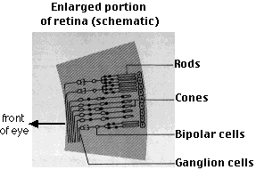
The visual receptors, rods and cones, synapse in the retina with several types of interneurons, e.g., bipolar cells.
The bipolar cells, in turn, synapse with ganglion cells.
The axons of ganglion cells make up the optic nerve and conduct impulses to the brain.
But each receptor does not have its own private circuit back to the brain. There are some 108 rods and cones in each eye, but only 106 ganglion cell axons in the optic nerve. Thus a single ganglion cell must receive inputs from a number of receptor cells.
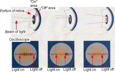
 By inserting an electrode in a single ganglion cell, it was shown (by Stephen W. Kuffler) that
Thus the optic nerve is not telling the brain simply that light has been detected but that contrast between light and dark (i.e., shape) has been detected.
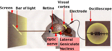
The axons of the optic nerve pass back close to the center of the base of the forebrain to a part of the diencephalon called the lateral geniculate nucleus (LGN), where they synapse with a new set of interneurons.

The axons from these cells lead up and back to the visual cortex in the occipital lobes.
Two associates of Kuffler, David H. Hubel and Torsten N. Wiesel inserted electrodes in these areas but instead of directing light into the eye, they projected images on a screen in front of the animal (an anesthetized cat or monkey).
Using this procedure, they found that
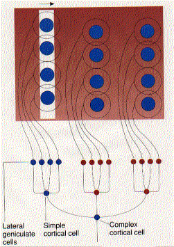
The preference that these first ("simple") cortical cells have for lines can be explained if we assume that they can be activated only if they receive inputs from a number of LGN cells whose areas of response (circular) are arranged in a line.
The diagram shows this mechanism by which the circular response areas of ganglion and LGN cells can be converted into the rectangular response areas found in the cells of the visual cortex.
Other cells ("complex cortical cells") still want their edges oriented in one direction, but the edges can now be moved across the screen. As the figure shows, this can be explained if a set of simple cortical cellsThus these complex cortical cells continue to respond to the stimulus even though its absolute position on the retina changes.
While these studies provide only the tiniest glimpse into the workings of the brain, they provide some clues of what will be found:The importance of these studies was recognized by the award of a Nobel Prize in 1981 to Hubel and Wiesel (too late for Kuffler, who died in 1980).
| Welcome&Next Search |