The central nervous system is made up of the
The spinal cord
- conducts sensory information from the peripheral nervous system (both somatic and autonomic) to the brain
- conducts motor information from the brain to our various effectors
- serves as a minor reflex center
The brain
- receives sensory input from the spinal cord as well as from its own nerves (e.g., olfactory and optic nerves)
- devotes most of its volume (and computational power) to processing its various sensory inputs and initiating appropriate — and coordinated — motor outputs.
- It is estimated that the brain contains about 86 billion neurons.
Both the spinal cord and the brain consist of
In the spinal cord, the white matter is at the surface, the gray matter inside.
In the brain of mammals, this pattern is reversed. However, the brains of "lower" vertebrates like fishes and amphibians have their white matter on the outside of their brain as well as their spinal cord.
Both the spinal cord and brain are covered in three continuous sheets of connective tissue, the meninges. From outside in, these are the
- dura mater — pressed against the bony surface of the interior of the vertebrae and the cranium
- the arachnoid
- the pia mater
The region between the arachnoid and pia mater is filled with cerebrospinal fluid (CSF).
The cells of the central nervous system are bathed in a fluid, called cerebrospinal fluid (CSF), that differs from that serving as the interstitial fluid (ISF) of the cells in the rest of the body.
- Cerebrospinal fluid leaves the capillaries in the choroid plexus of the brain.
- It contains far less protein than "normal" because of the blood-brain barrier, a system of tight junctions between the endothelial cells of the capillaries. (This barrier creates problems in medicine as it prevents many therapeutic drugs from reaching the brain.)
- CSF flows uninterrupted throughout the central nervous system
- through the central cerebrospinal canal of the spinal cord and
- through an interconnected system of four ventricles in the brain.
CSF returns to the blood through lymphatic vessels draining the brain.
In mice, the flow of CSF increases by 60% when they are asleep. Perhaps one function of sleep is to provide the brain a way of removing potentially toxic metabolites accumulated during waking hours.

31 pairs of spinal nerves arise along the spinal cord. These are "mixed" nerves because each contain both sensory and motor axons. However, within the spinal column,
- all the sensory axons pass into the dorsal root ganglion where their cell bodies are located and then on into the spinal cord itself.
- all the motor axons pass into the ventral roots before uniting with the sensory axons to form the mixed nerves.
The spinal cord carries out two main functions:
- It connects a large part of the peripheral nervous system to the brain. Information (nerve impulses) reaching the spinal cord through sensory neurons are transmitted up into the brain. Signals arising in the motor areas of the brain travel back down the cord and leave in the motor neurons.
- The spinal cord also acts as a minor coordinating center responsible for some simple reflexes like the withdrawal reflex.
The interneurons carrying impulses to and from specific receptors and effectors are grouped together in spinal tracts.
Impulses reaching the spinal cord from the left side of the body eventually pass over to tracts running up to the right side of the brain and vice versa. In some cases this crossing over occurs as soon as the impulses enter the cord. In other cases, it does not take place until the tracts enter the brain itself.
 The brain of all vertebrates develops from three swellings at the anterior end of the neural tube of the embryo. From front to back these develop into the
- forebrain (also known as the prosencephalon — shown in light color)
- midbrain (mesencephalon — gray)
- hindbrain (rhombencephalon — dark color) The human brain is shown from behind so that the cerebellum can be seen.
The human brain receives nerve impulses from
- the spinal cord and
- 12 pairs of cranial nerves
- Some of the cranial nerves are "mixed", containing both sensory and motor axons
- Some, e.g., the optic and olfactory nerves (numbers I and II) contain sensory axons only
- Some, e.g. number III that controls eyeball muscles, contain motor axons only.
 The main structures of the hindbrain (rhombencephalon) are the
- medulla oblongata
- pons and
- cerebellum
The medulla looks like a swollen tip to the spinal cord. Nerve impulses arising here
- rhythmically stimulate the intercostal muscles and diaphragm — making breathing possible [More]
- regulate heartbeat
- regulate the diameter of arterioles thus adjusting blood flow. [More]
The neurons controlling breathing have mu (µ) receptors, the receptors to which opioids, like heroin, bind. This accounts for the suppressive effect of opiates on breathing. [Discussion] Destruction of the medulla causes instant death.
The pons seems to serve as a relay station carrying signals from various parts of the cerebral cortex to the cerebellum. Nerve impulses coming from the eyes, ears, and touch receptors are sent on the cerebellum. The pons also participates in the reflexes that regulate breathing.
The reticular formation is a region running through the middle of the hindbrain (and on into the midbrain). It receives sensory input (e.g., sound) from higher in the brain and passes these back up to the thalamus. The reticular formation is involved in sleep, arousal (and vomiting).
The cerebellum consists of two deeply-convoluted hemispheres. Although it represents only 10% of the weight of the brain, it contains some 80% of all the neurons in the central nervous system.
Its most clearly-understood function is to coordinate body movements. People with damage to their cerebellum are able to perceive the world as before and to contract their muscles, but their motions are jerky and uncoordinated.
So the cerebellum appears to be a center for learning motor skills (implicit memory). Laboratory studies have demonstrated both long-term potentiation (LTP) and long-term depression (LTD) in the cerebellum.
The midbrain (mesencephalon) occupies only a small region in humans (it is relatively much larger in "lower" vertebrates). We shall look at only three features:
- the reticular formation: collects input from higher brain centers and passes it on to motor neurons.
- the substantia nigra: helps "smooth" out body movements; damage to the substantia nigra causes Parkinson's disease.
- the ventral tegmental area (VTA): packed with dopamine-releasing neurons that
The VTA seems to be involved in pleasure: nicotine, amphetamines and cocaine bind to and activate its dopamine-releasing neurons and this may account — at least in part (see below)— for their addictive qualities.
The midbrain along with the medulla and pons are often referred to as the "brainstem".
The human forebrain (prosencephalon) is made up of
- a pair of large cerebral hemispheres, called the telencephalon. Because of crossing over of the spinal tracts, the left hemisphere of the forebrain deals with the right side of the body and vice versa.
- a group of structures located deep within the cerebrum, that make up the diencephalon.
We shall consider four of its structures: the
- Thalamus.
- All sensory input (except for olfaction) passes through these paired structures on the way up to the somatic-sensory regions of the cerebral cortex and then returns to them from there.
- signals from the cerebellum pass through them on the way to the motor areas of the cerebral cortex.
- Lateral geniculate nucleus (LGN). All signals entering the brain from each optic nerve enter a LGN and undergo some processing before moving on the various visual areas of the cerebral cortex.
- Hypothalamus.
- The seat of the autonomic nervous system. Damage to the hypothalamus is quickly fatal as the normal homeostasis of body temperature, blood chemistry, etc. goes out of control.
- The source of 8 hormones, two of which pass into the posterior lobe of the pituitary gland.
- Posterior lobe of the pituitary.
Receives
from the hypothalamus and releases them into the blood.
 Each hemisphere of the cerebrum is subdivided into four lobes visible from the outside:
- frontal
- parietal
- occipital
- temporal
Hidden beneath these regions of each cerebral cortex is
- an olfactory bulb; they receive input from the olfactory epithelia.
- a striatum; they receive input from the frontal lobes and also from the limbic system (below). At the base of each striatum is a
- nucleus accumbens (NA).

The pleasurable (and addictive) effects of amphetamines, cocaine, and perhaps other psychoactive drugs seem to depend on their producing increasing levels of dopamine at the synapses in the nucleus accumbens (as well as the VTA).
- a limbic system; they receives input from various association areas in the cerebral cortex and pass signals on to the nucleus accumbens.
Each limbic system is made up of a:
- hippocampus. It is essential for the formation of long-term memories.
- an amygdala
The amygdala appears to be a center of emotions (e.g., fear). It sends signals to the hypothalamus and medulla which can activate the flight or fight response of the autonomic nervous system.
In rats, at least, the amygdala contains receptors for
- vasopressin whose activation increases aggressiveness and other signs of the flight or fight response;
- oxytocin whose activation lessens the signs of stress.
The amygdala receives a rich supply of signals from the olfactory system, and this may account for the powerful effect that odor has on emotions (and evoking memories).
It is estimated that the human brain contains some 86 billion (8.6 x 10¹⁰) neurons averaging 10,000 synapses on each; that is, almost 1015 connections. How to unravel the workings of such a complex system?
Several methods have been useful.
Microscopic examination with the aid of selective stains has revealed many of the physical connections created by axons in the brain.
This device measures electrical activity (brain "waves") that can be detected at the surface of the scalp. It can distinguish between, for example, sleep and excitement. It is also useful in diagnosing brain disorders such as a tendency to epileptic seizures.
Many cases of brain damage from, for example,
- strokes (interruption of blood flow to a part of the brain)
- tumors in the brain
- mechanical damage (e.g., bullet wounds)
have provided important insights into the functions of various parts of the brain.
Example 1:
Battlefield injury to the left temporal lobe of the cerebrum interferes with speech.
Example 2: Phineas P. Gage

In 1848, an accidental explosion drove a metal bar completely through the frontal lobes of Phineas P. Gage. Not only did he survive the accident, he never even lost consciousness or any of the clearly-defined functions of the brain. However, over the ensuing years, he underwent a marked change in personality. Formerly described as a reasonable, sober, conscientious person, he became — in the words of those observing him — "thoughtless, irresponsible, fitful, obstinate, and profane". In short, his personality had changed, but his vision, hearing, other sensations, speech, and body coordination were unimpaired. (Similar personality changes have since been often observed in people with injuries to their prefrontal cortex.)
The photograph (courtesy of the Warren Anatomical Museum, Harvard University Medical School) shows Gage's skull where the bar entered (left) and exited (right) in the accident (which occurred 12 years before he died of natural causes in 1861).
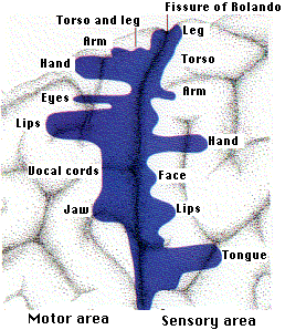
There are no pain receptors on the surface of the brain, and some humans undergoing brain surgery have volunteered to have their exposed brain stimulated with electrodes during surgery. When not under general anesthesia, they can even report their sensations to the experimenter.
Experiments of this sort have revealed a band of cortex running parallel to and just in front of the fissure of Rolando that controls the contraction of skeletal muscles. Stimulation of tiny spots within this motor area causes contraction of the muscles.
The area of motor cortex controlling a body part is not proportional to the size of that part but is proportional to the number of motor neurons running to it. The more motor neurons that activate a structure, the more precisely it can be controlled. Thus the areas of the motor cortex controlling the hands and lips are much larger than those controlling the muscles of the torso and legs.
A similar region is located in a parallel band of cortex just behind the fissure of Rolando. This region is concerned with sensation from the various parts of the body. When spots in this sensory area are stimulated, the patient reports sensations in a specific area of the body. A map can be made based on these reports.
When portions of the occipital lobe are stimulated electrically, the patient reports light. However, this region is also needed for associations to be made with what is seen. Damage to regions in the occipital lobe results in the person's being perfectly able to see objects but incapable of recognizing them.
The centers of hearing — and understanding what is heard — are located in the temporal lobes.
This is an imaging technique that uses a series of x-ray exposures taken from different angles. Computer software can integrate these to produce a three-dimensional picture of the brain (or other body region). CT scanning is routinely used to quickly diagnose strokes.
This imaging technique requires that the subject be injected with a radioisotope that emits positrons.

- water labeled with oxygen-15 (H215O) is used to measure changes in blood flow (which increases in parts of the brain that are active). The short half-life of 15O (2 minutes) makes it safe to use.
- deoxyglucose labeled with fluorine-18. The brain has a voracious appetite for glucose (although representing only ~2% of our body weight, the brain receives ~15% of the blood pumped by the heart and consumes ~20% of the energy produced by cellular respiration when we are at rest). When supplied with deoxyglucose, the cells are tricked into taking in this related molecule and phosphorylating it in the first step of glycolysis. But no further processing occurs so it accumulates in the cell. By coupling a short-lived radioactive isotope like 18F to the deoxyglucose and using a PET scanner, it is possible to visualize active regions of the brain.
The images above (courtesy of Michael E. Phelps from Science 211:445, 1981) were produced in a PET scanner. The dark areas are regions of high metabolic activity. Note how the metabolism of the occipital lobes (arrows) increases when visual stimuli are received.

Similarly, sounds increase the rate of deoxyglucose uptake in the speech areas of the temporal lobe.
The image on the right (courtesy of Gary H. Duncan from Talbot, J. D., et. al., Science 251: 1355, 1991) shows activation of the cerebral cortex by a hot probe (which the subjects describe as painful) applied to the forearm (which forearm?).
Most cancers consume large amounts of glucose (cellular respiration is less efficient than in normal cells so they must rely more on the inefficient process of glycolysis). Therefore PET scanning with 18F-fluorodeoxyglucose is commonly used to monitor both the primary tumor and any metastases.
This imaging technique uses powerful magnets to detect magnetic molecules within the body. These can be endogenous molecules or magnetic substances injected into a vein.
fMRI = Functional Magnetic Resonance Imaging
fMRI exploits the changes in the magnetic properties of hemoglobin as it carries oxygen. Activation of a part of the brain increases oxygen levels there increasing the ratio of oxyhemoglobin to deoxyhemoglobin.
The probable mechanism:
- The increased demand for neurotransmitters must be met by increased production of ATP.
- Although this consumes oxygen (needed for cellular respiration),
- it also increases the blood flow to the area.
- So there is an increase — not a decrease — in the oxygen supply to the region, which provides the signal detected by fMRI.
MEG detects the tiny magnetic fields created as individual neurons "fire" within the brain. It can pinpoint the active region with a millimeter, and can follow the movement of brain activity as it travels from region to region within the brain.
MEG is noninvasive requiring only that the subject's head lie within a helmet containing the magnetic sensors.
26 April 2023