- the mineralocorticoid aldosterone
- estrogens
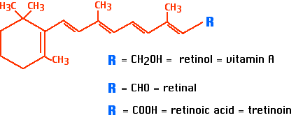
Steroid hormone receptors are proteins that have a binding site for a particular steroid molecule. Their response elements are DNA sequences that are bound by the complex of the steroid bound to its receptor.
The response element is part of the promoter of a gene. Binding by the receptor activates or represses, as the case may be, the gene controlled by that promoter.
It is through this mechanism that steroid hormones turn genes on (or off).
| Link to general discussion of eukaryotic promoters and the transcription factors that control them. |
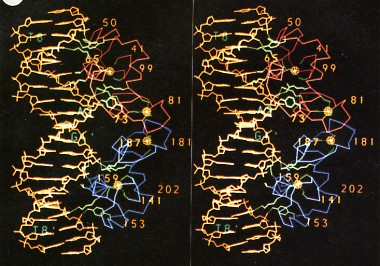
This image (courtesy of P. B. Sigler) shows a stereoscopic view of the glucocorticoid response element (DNA, the double helix shown in yellow at the left of each panel) with the glucocorticoid receptor (a protein homodimer, right portion of each panel) bound to it.
The DNA sequence of the glucocorticoid response element is
5' AGAACAnnnTGTTCT 3'
3' TCTTGTnnnACAAGA 5'
where n represents any nucleotide. (Note the inverted repeats.)
The glucocorticoid receptor, like all steroid hormone receptors, is a zinc-finger transcription factor; the zinc atoms are the four yellow spheres. Each is attached to four cysteines.
For a steroid hormone to regulate (turn on or off) gene transcription, its receptor must:Each of these functions depend upon a particular region of the protein (e.g., the zinc fingers for binding DNA). Mutations in any one region may upset the function of that region without necessarily interfering with other functions of the receptor.
Some of the hundreds of glucocorticoid response elements in the human genome activate gene transcription when bound by the hormone/receptor complex. Others inhibit gene transcription when bound by the hormone/receptor complex.
Example: When the stress hormone cortisol — bound to its receptor — enters the nucleus of a liver cell, the complex binds toNote that every type of cell in the body contains the same response elements in its genome. What determines if a given cell responds to the arrival of a hormone depends on the presence of the hormone's receptor in the cell.
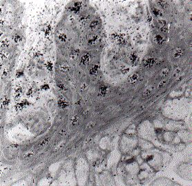
This autoradiograph (courtesy of Madhabananda Sar and Walter E. Stumpf) shows the endometrial cells from the uterus of a guinea pig 15 minutes after an injection of radioactive progesterone. The radioactivity has concentrated within the nuclei of the endometrial cells as shown by the dark grains superimposed on the images of the nuclei. The same effect is seen when radioactive estrogens are administered.
The cells of the endometrium are target cells for both progesterone and estrogens, preparing the uterus for possible pregnancy. [Link to discussion]
Nontarget cells (e.g. liver cells or lymphocytes) show no accumulation of female sex hormones. Although their DNA contains the response elements, their cells do not have the protein receptors needed.
| Welcome&Next Search |