The Immunological Synapse
Immunological responses, both
involve close contact between a T cell and an antigen-presenting cell (APC).
 Examples:
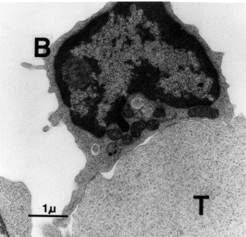
Electron microscopy (right) and several types of light microscopy have shown that these interactions involve a tight union between the two types of cells. This has been named the immunological synapse in recognition of the several ways in which it resembles the synapses of the nervous system.
The electron micrograph (courtesy of J. W. Uhr) shows a B cell and T cell bound to each other. The bar = 1 μm.
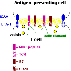
A number of pairs of molecules participate in the formation of the immunological synapse.
TCR/MHC-peptide pair
- On the T cell, the T-cell receptor for antigen (TCR) bound to
- a major histocompatibility complex (MHC) molecule on the antigen-presenting cell (APC).
- For CD4+ T cells, the MHC molecules are class II, and the binding is aided by CD4. [View]
- For CD8+ T cells (e.g., CTLs), the MHC molecules are class I, and the binding is aided by CD8. [View]
- In both cases, the MHC molecule has an antigenic peptide nestled in an exterior groove (MHC-peptide). [View]
- The TCR molecules are tethered by actin filaments in the cytoplasm.
- Several hundred TCR/MHC-peptide pairs are needed to stimulate a naive T cell to begin mitosis, but only 50 or so are needed to activate a "memory" T cell to do its work; that is, to become an effector T cell.
Costimulatory pairs
such as
- The costimulatory molecule CD28 on the T cell bound to
- its ligand, B7, on the APC
and
- the costimulatory molecules CD40L bound in the immunological synapse to
- CD40.
- Leukocyte Function-associated Antigen-1 (LFA-1), an integrin on the T cell, bound to
- InterCellular Adehesion Molecule-1 (ICAM-1) on the APC.
Cytokine receptors
Cytokine receptors also cluster in the synapse (not shown in the diagram) where they are exposed to cytokines secreted into the synapse.
Formation of an immunological synapse causes the T cell to
- become activated with various signal pathways turning on new gene transcription;
- release, by exocytosis, the contents of its vesicles:
- Type 1 helper T cells (Th1): lymphokines like IFN-γ and TNF-β [Link]
- Type 2 helper T cells (Th2): lymphokines like IL-4, IL-5, IL-10, and IL-13 that stimulate B cells [Link]
- Cytotoxic T Lymphocytes (CTLs): cytotoxic molecules like perforin and granzymes that kill the target. [Link]
B cells can also form an immunological synapse
While B cells can bind soluble antigens with their antigen receptors (BCRs), they can also bind antigens attached to the surface of dendritic cells. In doing so, they form an immunological synapse similar to that of B cell/T cell synapse.
Why an immunological synapse?
The molecules released by effector T cells (e.g., interleukins, perforin) are potent cytokines. Confining them to the immunological synapse ensures that they will not act nonspecifically against innocent bystander cells.
As for B cells and antigen-presenting dendritic cells, synapse formation appears to be a mechanism to cluster the antigens and BCRs in a small area, which enhances activation of the B cell.
Antigen-presenting cells like
can also present antigen to T cells by means of exosomes. These are tiny membrane-enclosed vesicles released by the cell. Their surface is studded with MHC-peptide complexes [View].
Antigen-presentation by exosomes may in some cases inhibit — rather than stimulate — an immune response.
9 November 2024
 Examples:
 Examples:
